


A Case of Endovascular Treatment for M2 Segment Occlusion of the Right Middle Cerebral Artery
Date:2025-05-15Category:Case ReportsViews:1296
Zenith devices used in this case:
Patient Information
- Female, 72 years old
- left extremities weakness for approximately 8 hours
- Left upper extremity: 0/5 strength,left lower extremity limb: 2/5 strength
- NIHSS score: 14 points
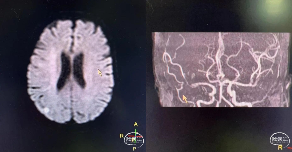
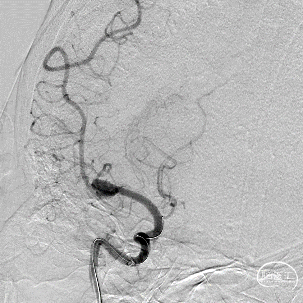
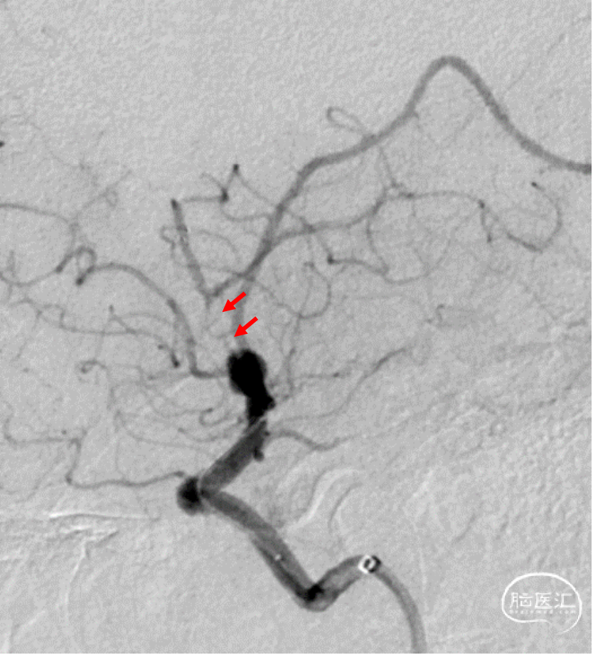
Preoperative Angiography
MRI from another hospital showed watershed infarction in the right hemisphere and occlusion of the right middle cerebral artery (M2 segment).
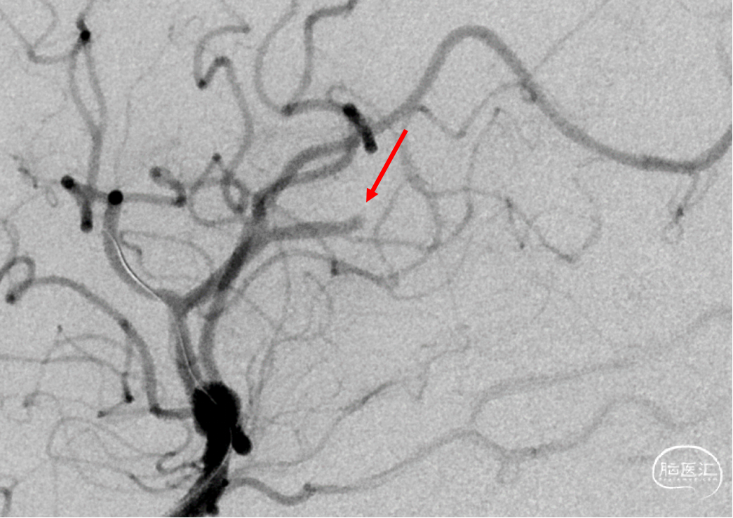
Frontal view angiography showed complete occlusion of the M2 superior trunk without a residual stump.
No residual segment was observed even after adjusting the projection angle.
A microwire was carefully advanced through the occlusion to locate the true lumen.
"Smoking" sign observed with the Rebar-18 microcatheter further confirmed entry into the true lumen.
A 4-20 stent retriever was deployed, but angiography showed poor proximal opacification, suggesting stenosis or residual thrombus.

Devices
- 8F Guiding Catheter
- Synchro Microguide Wire
- Rebar 18 Microcatheter
- XT 27 Microcatheter
- 5F Zenith Distal Access Catheter
- Solitaire FR 420 Stent Retriever
Procedure
Severe proximal M2 stenosis?
XT27 microcatheter was used to retrieve the stent.
The thrombus migrated distally, confirming embolic occlusion.
The occlusion site was a common location for M2 segment thromboembolism.
A second 4-20 stent retriever was deployed.

Under stent traction, a 5F Zenith Distal Access Catheter successfully advanced into the M2 superior trunk, enabling a combined aspiration and stent retrieval approach.
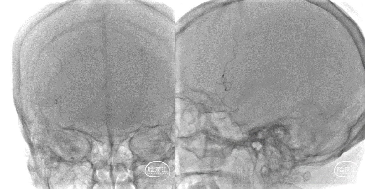
Postoperative Angiography
Frontal angiography showed excellent recanalization.
Lateral angiography also confirmed successful reperfusion.
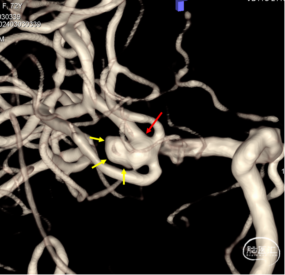
3D reconstruction revealed a classic bifurcation aneurysm with the M2 superior trunk originating from the aneurysm neck.
Postoperative CT and DWI
Surgeon Information
Bing Yang, The First Affiliate Hospital of Jinan University